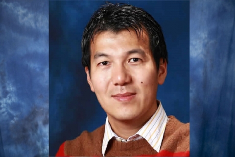
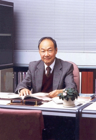
中大四學者 獲優秀青年科學家基金

今年首次接受香港及澳門年輕大學學者申請的「優秀青年科學家基金項目」,中文大學四名年輕科學家獲得本年度國家優秀青年科學基金,每人將獲一百三十萬元人民幣的資助,並容許在香港進行研究,資助期限三年。

Date: 
Wednesday, October 30, 2019
Media: 
Sing Tao

中大四學者獲國家優秀青年科學家基金

國家自然科學基金的「優秀青年科學家基金項目」,今年首次接受香港及澳門的年輕大學學者申請,香港中文大學四位年輕科學家呂愛蘭、田小雨、黃曦及盧怡君獲選,每人將獲得130萬元人民幣的資助,資助期限三年,容許在香港就其自選的研究方向進一步開展創新研究。

Date: 
Wednesday, October 30, 2019
Media: 
Ta Kung

Harvesting Energy from Walking Human Body

Date: 
2019-10-30
Thumbnail: 
Body: 

A research team led by Professor Wei-Hsin Liao from the Department of Mechanical and Automation Engineering has developed a lightweight smart materials-based energy harvester for scavenging energy from human motion, generating inexhaustible and sustainable power supply just from walking. Specifically, the device can capture biomechanical energy from the motion of the human knee and then convert it to electricity which can be used to power wearable electronics such as pedometers, health monitors, and GPS. This work has been published in Applied Physics Letters and recommended as a featured article by editors.

 
So far, researchers have developed large devices to use human motion for generating electricity, such as electromagnetic generator-based energy harvesters for capturing energy when people are walking on treadmills or riding bicycles. However, these bulky devices hamper the users’ locomotion and at the same time increase their burden, because of the considerable weight and large interaction force between the harvesters and the human body. This considerably restricts the wide use of these devices. To overcome that, a research team led by Professor Liao proposed and developed a lightweight energy harvester employing piezoelectric macro fiber composites integrated with novel mechanical structures.
 
Piezoelectric macro fiber composites are lightweight materials, which can produce electricity under deformation. The proposed energy harvester employs a bending beam and a slider-crank mechanism to capture the motion of the human knee when walking. Then, the captured motion is used to deform piezoelectric macro fiber composites pieces bonded to the bending beam so that electricity is produced when the human knee flexes or extends. 
 
Professor Liao said, “The human knee joint has a larger range of motion than other lower limb joints such as the ankle and hip, which enables energy harvesters to capture the motion more easily and generate more electricity.” The prototype harvester, made by piezoelectric macro fiber composites, can generate an average power of 1.6 mW, when the wearer walks at about 2-6.5 km/h. The generated electricity is efficient to power common wearable electronic devices such as smart bands. Furthermore, the prototype weighs only 307 grams. When walking with it, the wearer’s metabolic cost is almost the same as that when walking without the device. Unlike the existing electromagnetic generator-based energy harvesters, the lightweight smart materials-based energy harvester can capture energy from human motion without increasing the wearer’s burden. It is expected to significantly promote the use of biomechanical energy harvesters.
 
Professor Liao stated, “This apparatus will attract much attention from mountaineers and hikers. If they get lost in remote mountains or a wilderness where the power grid is unavailable, the device can derive energy from their motion and convert it to electricity, enabling wearers to continuously monitor their vital signs, know their position, or even send out an SOS signal at any time when they need help. At present, we are focusing on improvement in the performance of the harvester by reducing the weight of the device and increasing energy harvesting efficiency. We plan to commercialise the harvester and market it through cooperating with garment manufacturers to embed the device in sportswear.”
The smart material of the device will bend alongside the motion of the human knee, and convert the biomechanical energy to electricity.

Professor Liao (right) and his Postdoctoral fellow Gao Fei (left) develop this energy harvester device in six months’ time.

Professor Liao

The energy harvester device is extremely light with only 307 grams.


Filter: Dept: 
Faculty
MAE
Media Release

中大新掃描技術助生物醫學研究 花粉3D成像只需0.55秒

神經活動一般在10毫秒量級時間內完成,傳統顯微鏡難以直接觀察現象。香港中文大學工程學院開發出高速成像方法,結合壓縮感知演算法與數碼全息顯微鏡,可在1秒內完成3D樣品的雙光子螢光成像,比傳統點掃描方法快3至5倍。

Date: 
Friday, October 25, 2019
Media: 
HKET

中大開發新成像方法促生物醫學研究

香港中文大學工程學院機械與自動化工程學系陳世祈教授及其團隊,結合壓縮感知演算法與數碼全息顯微鏡,開發了一種高速成像方法,在1 秒內即可完成對三維樣品的雙光子螢光成像,速度是傳統點掃描方法的3 至5 倍。相關研究成果近日於著名期刊《Optics Letters》發表。

Date: 
Friday, October 25, 2019
Media: 
Hong Kong Commercial Daily

中大研發高速三維成像法

人們過往觀察生物中數百個神經元的活動時,往往因速度不夠快,難以從傳統顯微鏡中直接觀察這些現象,故中文大學機械與自動化工程學系教授陳世祈帶領其研究團隊,結合「壓縮感知演算法」及數碼全息顯微鏡,開發出一種高速成像方法,能於一秒內完成對三維樣品的雙光子熒光成像,解決了數字微鏡器件無法調製超快雷射的問題,這項研究更發表於著名期刊《Optics Letters》。

Date: 
Friday, October 25, 2019
Media: 
Sing Tao

活體高速3D成像中大助研究光遺傳學

香港文匯報訊(記者高鈺)快速的生物成像方法,對生命科學的探索至關重要。中文大學機械與自動化工程學系副教授陳世祈及其團隊,結合壓縮感知演算法與數碼全息顯微鏡,成功開發一種高速雙光子熒光成像法,在1秒內即可完成活體3D成像,速度是傳統點掃描方法的3 倍至5倍。新技術可望對神經系統及光遺傳學研究帶來貢獻,促進生物醫學領域發展。

Date: 
Friday, October 25, 2019
Media: 
Wen Wei

Novel Imaging Approach Bringing Faster 3D Imaging for Biomedical Researches

Date: 
2019-10-24
Thumbnail: 
Body: 

By combining a compressive sensing algorithm with a digital holographic microscope, Prof. Shih-Chi CHEN of the Department of Mechanical and Automation Engineering, Faculty of Engineering and his research team have developed a high-speed imaging method. The new approach is able to produce two-photon microscopy images of a 3D sample in one second, which is at a speed three to five times that of the conventional point-scanning method. The research result has been published in the journal Optics Letters recently.

Activities of neurons are generally completed on a time scale of 10 milliseconds, which makes it hard for conventional microscopes to observe these phenomena directly. This new compressive sensing two-photon microscopy can be applied to 3D imaging of the nerve distribution of living things or to monitoring activities from hundreds of neurons simultaneously.
 
New multi-focus laser scanning method to break the scanning speed limit of two-photon microscope
 
Two-photon microscopy works by delivering ultrafast pulses of infrared laser light to the sample, where it interacts with fluorescent labels to create an image. It is extensively used for biological researches because of its ability to produce high-resolution 3D images up to a depth of one millimeter in a living tissue. These advantages, however, come with a limited imaging speed of the two-photon microscopy because of the weak fluorescent signal.
 
To speed up scanning, the research team developed a multi-focus laser illumination method that uses a digital micromirror device (DMD). The research solves the problem of conventional DMD being unusable to work with ultrafast laser, enabling them to be integrated and used in beam shaping, pulse shaping, and two-photon imaging.
 
The DMD generates 30 points of focused laser light on randomly selected locations within a specimen. The position and intensity of each point of light are controlled by a binary hologram that is projected onto the device. During each measurement, the DMD reflashes the hologram to change the position of each focus and records the intensity of the two-photon fluorescence with a single-pixel detector. Although, in many ways, the DMD multi-focus scanning is more flexible and faster than traditional mechanical scanning, the speed is still limited by the DMD’s refresh rate.
 
Combining the compressive sensing algorithm to further improve the imaging speed
 
The researchers further increased the imaging speed in this research by combining multi-focus scanning with compressive sensing. This approach enables image acquisition with fewer measurements. This is because it carries out image measurement and compression in a single step and then uses an algorithm to rebuild the images from the measurement results. For two-photon microscopy, it can reduce the number of measurements by between 70% and 90%.
 
After conducting a simulation experiment to demonstrate the new method’s performance and parameters, the researchers tested it with two-photon imaging experiments. These experiments demonstrated the technique’s ability to produce high-quality 3D images with high imaging speeds from any field of view. For example, they were able to acquire 3D images from a pollen grain, in just 0.55 seconds. The same images acquired with traditional point scanning took 2.2 seconds. 
 
Prof. Shih-Chi Chen said, “This method achieved a three to five times enhancement in imaging speed without sacrificing the resolution. We believe this novel approach will lead to new discoveries in biology and medicine, such as optogenetics. The team is now working to further improve the speed of the reconstruction algorithm and image quality. We also plan to use the DMD together with other advanced imaging techniques, which allows imaging in deeper tissues.”
 

Prof. Shih-Chi CHEN (1st from right, front row) and his research team.

The researchers prepared two-photon microscopy images of a pollen grain by using (a) traditional point-scanning and (b) the new compressive imaging approach. The point-scanning imaging time was 2.2 seconds while the compressive imaging time required only 0.55 seconds.

Researchers have developed a system that increases the imaging speed of two-photon microscopy up to three to five times without compromising resolution. On the left is a CAD model of the compact, custom built, two-photon microscopy system. The free-space optics inside are pictured on the right.


Filter: Dept: 
Faculty
MAE

中大研納米機械人 體內遙控攜藥治中風 五毫米血塊 20分鐘溶解

中風是本港第四大致命疾病,每年逾三千人死亡,現時常見的治療方法具一定風險,中文大學機械與自動化工程學系教授張立,把氧化鐵納米微粒移接到螺旋藻的表面,研發可攜帶藥物的磁性生物合成納米機械人,能以磁場控制其移動到目標部位施藥,納米機械人只需二十分鐘,便可溶解五毫米的血塊,成為無綫操控微型醫療機械人技術的里程碑。

Date: 
Tuesday, October 22, 2019
Media: 
SING TAO

中大研發評估納米發電新技術

納米發電機透過收集熱能、機械能等轉化為電能,它的出現為小型電子設備提供持續穩定供電,功能亦愈趨多元化。香港

Date: 
Tuesday, October 15, 2019
Media: 
Hong Kong Commercial Daily

Pages